Suite à une grève chez bpost il n'est temporairement pas possible de choisir pour livraison à domicile ou à une autre adresse. Besoin de quelque chose en urgence ? Choissisez pour retrait en magasin ou passez plutôt dans un magasin Club à proximité.
- Retrait gratuit dans votre magasin Club
- 7.000.000 titres dans notre catalogue
- Payer en toute sécurité
- Toujours un magasin près de chez vous
Suite à une grève chez bpost il n'est temporairement pas possible de choisir pour livraison à domicile ou à une autre adresse. Besoin de quelque chose en urgence ? Choissisez pour retrait en magasin ou passez plutôt dans un magasin Club à proximité.
- Retrait gratuit dans votre magasin Club
- 7.000.0000 titres dans notre catalogue
- Payer en toute sécurité
- Toujours un magasin près de chez vous


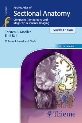
Pocket Atlas of Sectional Anatomy, Volume I: Head and Neck
Computed Tomography and Magnetic Resonance Imaging
Torsten Bert Möller, Emil Reif
44,45 €
+ 88 points
Description
This comprehensive, easy-to-consult pocket atlas is renowned for its superb illustrations and ability to depict sectional anatomy in every plane. Together with its two companion volumes, it provides a highly specialized navigational tool for all clinicians who need to master radiologic anatomy and accurately interpret CT and MR images.
Special features of Pocket Atlas of Sectional Anatomy
- Didactic organization in two-page units, with high-quality radiographs on one side and brilliant, full-color diagrams on the other
- Hundreds of high-resolution CT and MR images made with the latest generation of scanners (e.g., 3T MRI, 64-slice CT)
- Consistent color coding, making it easy to identify similar structures across several slices
- Concise, easy-to-read labeling of all figures
Updates for the 4th edition of Volume I:
- New cranial CT imaging sequences of the axial and coronal temporal bone
- Expanded MR section, with all new 3T MR images of the temporal lobe and hippocampus, basilar artery, cranial nerves, cavernous sinus, and more
- New arterial MR angiography sequences of the neck and additional larynx images
Compact, easy-to-use, highly visual, and designed for quick recall, this book is ideal for use in both the clinical and study settings.
Spécifications
Parties prenantes
- Auteur(s) :
- Editeur:
Contenu
- Nombre de pages :
- 333
- Langue:
- Anglais
- Collection :
Caractéristiques
- EAN:
- 9783131255044
- Date de parution :
- 11-12-13
- Format:
- Livre broché
- Format numérique:
- Trade paperback (VS)
- Dimensions :
- 130 mm x 189 mm
- Poids :
- 453 g