- Retrait gratuit dans votre magasin Club
- 7.000.000 titres dans notre catalogue
- Payer en toute sécurité
- Toujours un magasin près de chez vous
- Retrait gratuit dans votre magasin Club
- 7.000.0000 titres dans notre catalogue
- Payer en toute sécurité
- Toujours un magasin près de chez vous
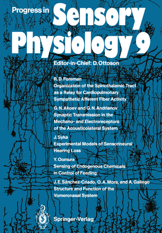
Progress in Sensory Physiology 9
150,48 €
+ 300 points
Description
Sympathetic afferent fibers originate from a visceral organ, course in the thoracolumbar rami communicantes, have cell bodies located in dorsal root ganglia, and terminate in the gray matter of the spinal cord. Sympathetic afferent fibers from the heart transmit information about noxious stimuli associated with myocardial ischemia, i. e. angina pectoris. Previous reviews have described the characteristics of cardiovascular sympathetic afferent fibers (Bishop et al. 1983; Malliani 1982). This review summarizes that work and focuses on the neural mechanisms underlying the complexities of angina pectoris. In order to understand anginal pain, cells forming the classical pain pathway, the spinothalamic tract (STn, were chosen for study. These cells were chosen to address questions about anginal pain because they transmit nociceptive informa- of pain. Antidromic tion to brain regions that are involved in the perception activation of STT cells provided a means of identifying cells involved with trans- mission of nociceptive information in anesthetized animals. Other ascending pathways may also transmit nociceptive information, but many studies show that the STT plays an important role. Visceral pain is commonly referred to overlying somatic structures. The pain of angina pectoris can be sensed over a wide area of the thorax: in the retrosternal, precordial anterior thoracic, and anterior cervical regions of the chest; in the left or sometimes even the right shoulder, arm, wrist, or hand; or in the jaw and teeth (Harrison and Reeves 1968).
Spécifications
Parties prenantes
- Editeur:
Contenu
- Nombre de pages :
- 227
- Langue:
- Anglais
- Collection :
- Tome:
- n° 9
Caractéristiques
- EAN:
- 9783642740602
- Date de parution :
- 16-12-11
- Format:
- Livre broché
- Format numérique:
- Trade paperback (VS)
- Dimensions :
- 170 mm x 244 mm
- Poids :
- 381 g

Les avis
Nous publions uniquement les avis qui respectent les conditions requises. Consultez nos conditions pour les avis.


